brain mri
Tired of deploying image evaluation scripts on your local machine? ASL image evaluation for brain perfusion analysis can be as easy as this: click, click.
Watch this video to see how images are uploaded from your local network to the central mTRIAL server – pseudonymization on the fly and image categorizaton included.

World Multiple Sclerosis Day, as a day of global solidarity, emphasizes the need to raise awareness of this disease, improve diagnosis and treatment, and fund research to curb multiple sclerosis.
At mediri we develop and refine structural analysis methods to support the diagnosis and follow-up of multiple sclerosis.
An important imaging biomarker for MS is smouldering lesions. Their AI-aided detection on MR images also makes disease activity visible that cannot be seen with the naked eye.

You want the brain MRIs of your MS patients to be evaluated by the SPM Lesion Segmentation Tool but without installing and adapting any software packages? Or do you need consistent evaluations across multiple centres of a clinical trial? mTRIAL offers Lesion analysis of FLAIR images based on the SPM LST algorithm and produces clear reports. You only need to upload your image data to the mTRIAL platform, pseudonymization is performed on the fly, evaluation and reporting can be done within minutes.
Interested? Don’t hesitate to contact us.
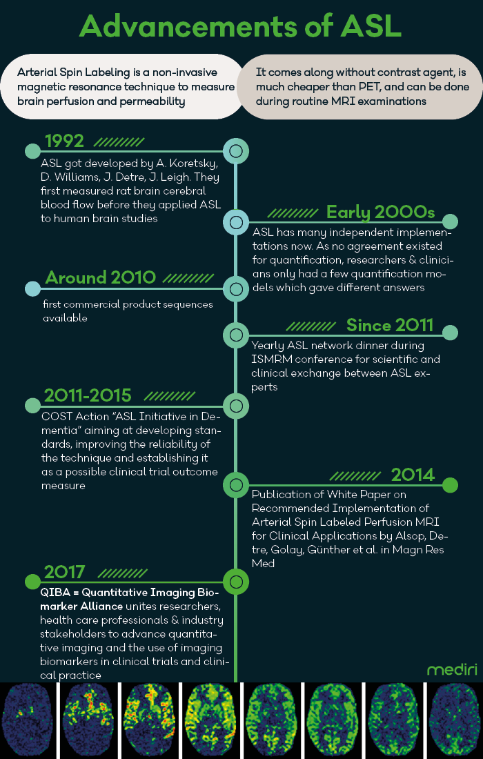
30 years after the first successful Arterial Spin Labeling (ASL) MR image acquisition to measure cerebral blood flow in rat brain, the early detection of Alzheimer’s using ASL gains enormous importance with the recent publication of the study results for Lecanemab. Now that there is hope for efficient therapies in dementia diseases, it is a good time to review the steps that have been gone since then. Many initiatives aiming at reliable and comparable quantification of brain perfusion measurements are paving the way for broad usage of ASL in clinical studies focusing on Alzheimer‘s and other dementia diseases.