Research

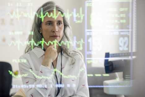
Our focus lies on a user-friendly display of ASL evaluation results to support efficient study workflows and reliable results. We provide interactive reports for quick overview of a participant as well as pdf reports for structured archiving.


An aortic aneurysm or even an aortic dissection is often discovered as an incidental finding on abdominal CT scans. In case of doubt, however, the patient must be treated as quickly as possible. We are developing an automatic, AI-supported evaluation of all abdominal CT images in the cloud, which alerts the physician on duty directly in the event of a critical finding, so that the patient can be prioritised.

The likelihood of breast cancer recurrence is still difficult to predict. Together with nine partners in the EU, we are the first to combine radiological, histo-pathological and clinical data of patients in one model to predict relapse of distant metastases.
State of the art AI solutions for image and data analysis form the basis for our development in all areas of our IT products. Through active research, we ensure that we will continue to offer cutting edge solutions today and in the future. A selection of our research projects in the field of artificial intelligence:
ASPIRE: Alzheimer’s early detection using ASL (Arterial Spin Labelling)
KI4MS: AI-assisted detection of smouldering lesions for follow-up of MS
DEEPRAY: Automatic detection of aortic aneurysms

Analysis software for the non-invasive perfusion imaging technique Arterial Spin Labeling (ASL) is integrated in our professional solution “mTRIAL”, to support clinical researchers to conduct research and clinical trials in real-time. Current highlights are the new web-based DICOM viewer and the atlas based regional perfusion analysis, allowing automated analysis of longitudinal studies. Contact us if you want to learn more about brain perfusion analysis with ASL for clinical studies.
Our great project partners:
Goldstandardphantoms Ltd, established in London, UK, builds highest accuracy perfusion phantoms used for quality control, ensuring stable and reliable image quantification in clinical trials.
The Amsterdam University Medical Center in the Netherlands is at the forefront of clinical research in Alzheimer’s and dementia diseases. They boost the ASL community research by contributing their knowledge to the analysis tool “ExploreASL”.
The University of Applied Sciences Darmstadt, Germany, has a strong focus on computer vision technologies. They provide the AI for AIzheimer’s prediction based on the most advanced neural networks.

Biomarkers cover a broad spectrum: from body temperature and blood count to the expression of certain genes in cancer cells. MR-based biomarkers are particularly elegant because they are non-invasive, i.e., they do not require a biopsy or blood sample.

ASL has the potential to improve diagnosis and treatment monitoring in Alzheimer’s Disease, especially for older adults with cardiovascular risk factors.
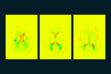
Structural changes caused by inflammatory processes offer signs of the future course of multiple sclerosis and also provide new information for other disease patterns. The tried and tested VGM algorithm visualises these changes and creates a “map” of the brain. The evaluation determines tissue changes with 100 million degrees of freedom.
Image: KI4KMU – Example of an AI-supported VGM evaluation, which was developed as part of a project funded by the Baden-Württemberg Ministry of Economics together with the University Medical Center Mannheim and MedicalSyn GmbH in Stuttgart.
We are working on identifying early-stage Alzheimer’s disease on MR images. Artificial intelligence helps us to recognise and assess specific patterns in the perfusion of the brain.