slides

An aortic aneurysm or even an aortic dissection is often discovered as an incidental finding on abdominal CT scans. In case of doubt, however, the patient must be treated as quickly as possible. We are developing an automatic, AI-supported evaluation of all abdominal CT images in the cloud, which alerts the physician on duty directly in the event of a critical finding, so that the patient can be prioritised.

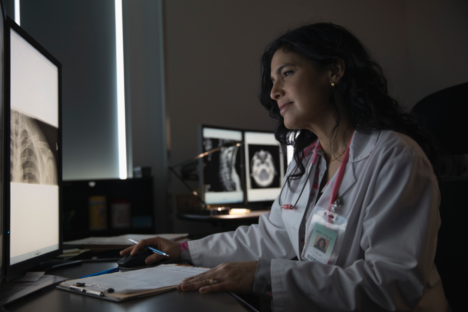
The likelihood of breast cancer recurrence is still difficult to predict. Together with nine partners in the EU, we are the first to combine radiological, histo-pathological and clinical data of patients in one model to predict relapse of distant metastases.

ULTRAWEAR is a portable ultrasound system that is used in physiotherapy for back pain. Our AI-supported real-time analysis helps patients perform their physiotherapy exercises correctly.
The system covers a wide range of ultrasound therapies: from hyperthermia to thermal ablation and cavitation. In this research project, mediri is working on crucial software components in the field of image registration and tracking.

CURE-OP is a new type of ultrasound therapy system for polytherapy of cancer. It enables the combined treatment with ultrasound and ionising radiation.
mediri heads the interdisciplinary research consortium that aims to make this treatment method easier and safer. In this project we develop the tracking of the catheter on ultrasound images during treatment.
Image source: „3D-XGuide: open-source X-ray navigation guidance system” by Ina Vernikouskaya et al. in Int.J. of Computer Assisted Radiology and Surgery (2020)

Surgical interventions in cardiology are increasingly being performed using minimally invasive methods. These include trans vascular interventions that use the vessels as access.
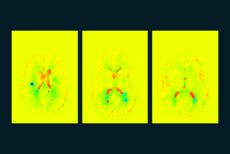
Structural changes caused by inflammatory processes offer signs of the future course of multiple sclerosis and also provide new information for other disease patterns. The tried and tested VGM algorithm visualises these changes and creates a “map” of the brain. The evaluation determines tissue changes with 100 million degrees of freedom.
Image: KI4KMU – Example of an AI-supported VGM evaluation, which was developed as part of a project funded by the Baden-Württemberg Ministry of Economics together with the University Medical Center Mannheim and MedicalSyn GmbH in Stuttgart.
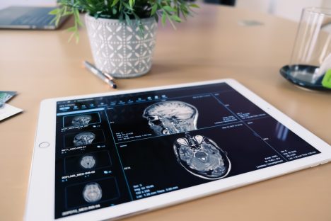
We are working on identifying early-stage Alzheimer’s disease on MR images. Artificial intelligence helps us to recognise and assess specific patterns in the perfusion of the brain.
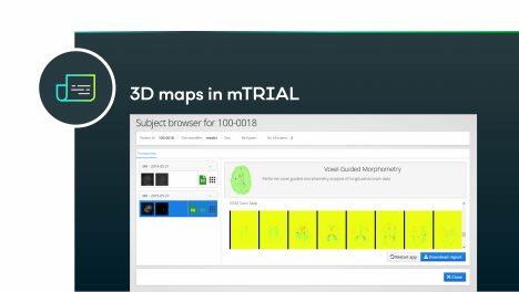
In the past this has been seen as a time-consuming process previously only used in academic settings. In the KI4MS project, we bring this valuable technology to the radiological practice. To do this, we use a neural network that has been specially trained for the structural changes. This artificial intelligence needs less than ten minutes to calculate a 3D map of the tissue changes.
Image: KI4KMU – Example of an AI-supported VGM evaluation, which was developed as part of a project funded by the Baden-Württemberg Ministry of Economics together with the University Medical Center Mannheim and MedicalSyn GmbH in Stuttgart.