Imaging biomarkers

Imaging Biomarkers – patient-friendly early warning systems

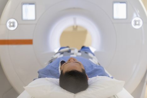
With imaging biomarkers, we use biological characteristics to monitor disease progression or support a diagnosis. The advantage is that they are usually non-invasive, do not require contrast media and can detect a disease even before it manifests.

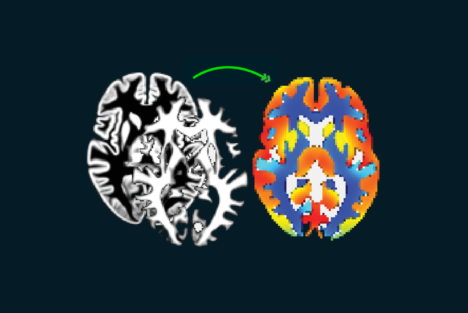
Analysis methods supported by AI technologies

Today, mediri uses AI-based technology in various research projects on imaging biomarkers. This state-of-the-art technology helps to develop and refine analysis methods.

A selection:

  • ASPIRE: Non-invasive Alzheimer’s early detection using ASL (Arterial Spin Labelling)
  • MS Atrophy: support of diagnosis and follow-up of multiple sclerosis
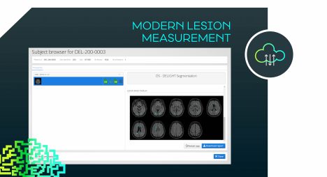
  • KI4MS: AI-assisted detection of smouldering lesions for follow-up of MS

Read more about the research projects in the slider below

Tracking Alzheimer’s non-invasively I – ASPIRE

Using arterial spin labelling (ASL), the blood flow in the brain is displayed non-invasively on MR images – without contrast media. As the head of a top-class European research consortium…

Non-invasively tracking Alzheimer’s II ­­­­– ASPIRE

… we establish a standardised workflow that facilitates the evaluation and development of this first ASL-based Alzheimer’s imaging biomarker.

Image source: „ExploreASL: An image processing pipeline for multi-center ASL perfusion MRI studies” by Henk J.M.M. Mutsaerts et al. in NeuroImage (2020)

Multiple sclerosis in our sight

We develop and refine structural analysis methods to support the diagnosis and follow-up of multiple sclerosis. Two examples of modern imaging biomarker analysis methods are given next:

Multiple sclerosis in our sight I – MS Atrophy

MS can manifest in irregular relapses of increased disease activity. Atrophy of the cervical spinal cord is a relatively new biomarker that correlates with disease activity and is used to monitor progression.

Image: MS Atrophy – Automatically segmented medulla of the spine for longitudinal analysis in mTRIAL Webapplication

Multiple Sclerosis in our Sight II – KI4MS

Another imaging biomarker for MS is smouldering lesions. Their AI-aided detection on MR images also makes disease activity visible that cannot be seen with the naked eye.

Image: KI4KMU – Example of an AI-supported VGM evaluation, which was developed as part of a project funded by the Baden-Württemberg Ministry of Economics together with the University Medical Center Mannheim and MedicalSyn GmbH in Stuttgart.

« Biomarkers cover a broad spectrum: from body temperature and blood count to the expression of certain genes in cancer cells. MR-based biomarkers are particularly elegant because they are non-invasive, i.e., they do not require a biopsy or blood sample. »

Prof. Dr. Matthias Günther,
CEO