Artificial intelligence

AI – When imaging thinks along

AI is changing the path of radiology services through a collection of algorithms and machine learning tools which support the medical professional. Cloud-based artificial intelligence works faster than any human radiologist, with the added capability to be in multiple places at the same time. In other words: artificial intelligence has the potential to improve medical care across the board for both patients and medical professionals. This can relieve doctors of repetitive, time-consuming tasks leaving more time for treating patients. In addition, AI brings particularly complex diagnostic processes into medical practices that were previously reserved for specialised departments.

AI – When imaging thinks along

State of the art AI solutions for image and data analysis form the basis for our development in all areas of our IT products. Through active research, we ensure that we will continue to offer cutting edge solutions today and in the future. A selection of our research projects in the field of artificial intelligence:
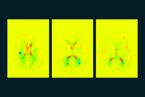
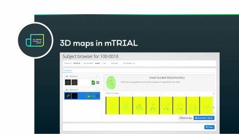
ASPIRE – Alzheimer’s early detection using ASL (Arterial Spin Labelling)

DELIGHT – Deep learning to quantify hyperintense lesions

KI4MS – Smouldering lesions in multiple sclerosis

TRANSNAV – Real-time tracking of organs

On the trail of Alzheimer’s – ASPIRE

We are working on identifying early-stage Alzheimer’s disease on MR images. Artificial intelligence helps us to recognise and assess specific patterns in the perfusion of the brain.

Modern lesion measurement for all practices I – DELIGHT

Measuring certain lesions in the brain enables effective stroke prevention. As does research into the influence of microangiopathy on cognition, dementia risk and the risk of stroke. However, visual inspection of high-resolution MR images is both time-consuming and a manual process.

Image: Automatically segmented white matter hyperintensities in the brain, shown in several slices

Modern lesion measurement for all practices II – DELIGHT

Using a dedicated AI approach, we enable fully automatic detection of inflammatory lesions, this within a few minutes after the MR scan. Integrated into mediri’s Cloud Platform, we provide the new artificial intelligence in all radiological practices with minimal effort.

Image: Results of analysis displayed in mTRIAL Webapplication

High-speed upgrade for brain mapper I – KI4MS

Structural changes caused by inflammatory processes offer signs of the future course of multiple sclerosis and also provide new information for other disease patterns. The tried and tested VGM algorithm visualises these changes and creates a “map” of the brain. The evaluation determines tissue changes with 100 million degrees of freedom.

Image: KI4KMU – Example of an AI-supported VGM evaluation, which was developed as part of a project funded by the Baden-Württemberg Ministry of Economics together with the University Medical Center Mannheim and MedicalSyn GmbH in Stuttgart.

High-speed upgrade for brain mapper II – KI4MS

In the past this has been seen as a time-consuming process previously only used in academic settings. In the KI4MS project, we bring this valuable technology to the radiological practice. To do this, we use a neural network that has been specially trained for the structural changes. This artificial intelligence needs less than ten minutes to calculate a 3D map of the tissue changes.

Image: KI4KMU – Example of an AI-supported VGM evaluation, which was developed as part of a project funded by the Baden-Württemberg Ministry of Economics together with the University Medical Center Mannheim and MedicalSyn GmbH in Stuttgart.

A look inside the patient – real-time image analysis

In the area of ​​real-time image analysis, we use modern AI algorithms such as recurrent neural networks or action recognition, too. Combined with our robust analysis method of particle filter-based tracking, the results of our technology meet every requirement put forth.

Image: Use cases of different projects. In the case of prostate detection (left image), the mediri tracking algorithm generates a vector field describing the movement of a delineated structure.

« Precise, individual diagnosis of dementia, even in smaller practices and clinics without a research background, is our vision. Artificial intelligence is predestined to support physicians in this work. »

Dr. Julia Müller,
COO