Research

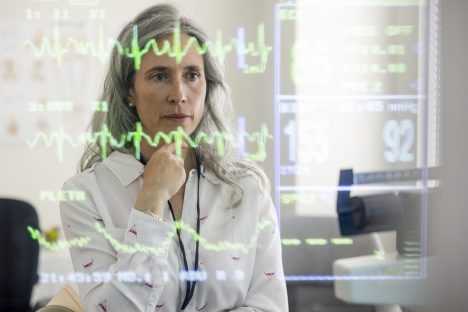
An aortic aneurysm or even an aortic dissection is often discovered as an incidental finding on abdominal CT scans. In case of doubt, however, the patient must be treated as quickly as possible. We are developing an automatic, AI-supported evaluation of all abdominal CT images in the cloud, which alerts the physician on duty directly in the event of a critical finding, so that the patient can be prioritised.

The likelihood of breast cancer recurrence is still difficult to predict. Together with nine partners in the EU, we are the first to combine radiological, histo-pathological and clinical data of patients in one model to predict relapse of distant metastases.
State of the art AI solutions for image and data analysis form the basis for our development in all areas of our IT products. Through active research, we ensure that we will continue to offer cutting edge solutions today and in the future. A selection of our research projects in the field of artificial intelligence:
ASPIRE: Alzheimer’s early detection using ASL (Arterial Spin Labelling)
KI4MS: AI-assisted detection of smouldering lesions for follow-up of MS
DEEPRAY: Automatic detection of aortic aneurysms
This technology makes it possible to display structures and changes in the body in real time. Physicians can theoretically “look directly into the patient” even during minimally invasive operations or therapies, which makes these interventions faster, easier and safer.
This technology is mainly used for therapy monitoring. For example, in catheter interventions or in the ablation of diseased tissue using focused ultrasound. We participate in various joint projects and are responsible for data analysis and the development of real-time AI for ultrasound, X-ray fluoroscopy and magnetic resonance tomography.
A selection of our research projects:
TRANSNAV: catheter tracking during cardiological interventions
CURE-OP: ultrasound therapy against cancer
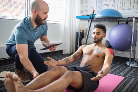
ULTRAWEAR: real-time support for physiotherapy
You can find more about our research projects with real-time image analysis in the slider:
ULTRAWEAR is a portable ultrasound system that is used in physiotherapy for back pain. Our AI-supported real-time analysis helps patients perform their physiotherapy exercises correctly.
The system covers a wide range of ultrasound therapies: from hyperthermia to thermal ablation and cavitation. In this research project, mediri is working on crucial software components in the field of image registration and tracking.

CURE-OP is a new type of ultrasound therapy system for polytherapy of cancer. It enables the combined treatment with ultrasound and ionising radiation.
mediri heads the interdisciplinary research consortium that aims to make this treatment method easier and safer. In this project we develop the tracking of the catheter on ultrasound images during treatment.
Image source: „3D-XGuide: open-source X-ray navigation guidance system” by Ina Vernikouskaya et al. in Int.J. of Computer Assisted Radiology and Surgery (2020)

Surgical interventions in cardiology are increasingly being performed using minimally invasive methods. These include trans vascular interventions that use the vessels as access.

Biomarkers cover a broad spectrum: from body temperature and blood count to the expression of certain genes in cancer cells. MR-based biomarkers are particularly elegant because they are non-invasive, i.e., they do not require a biopsy or blood sample.